Skip to content
North London, ON
South London, ON
(519) 641-1411
Location
South Location
North Location
Services
Cosmetic Dentistry
dental implants London Ontario
Dentures
Emergency Dentist London, Ontario
Family Dentistry
Fastbraces
Clear Aligners – A Modern Solution for Every Age
Laser Dentistry
Root Canal Treatment London
Sedation Dentistry
Teeth Cleaning
Teeth Whitening
Wisdom Teeth Extraction
About us
Special Offers
Patients
Video Testimonial
Patient Gallery
Blogs
Contact Us
South Location
North Location
Tag: Digital
Your Dentist in London, Ontario - Apple Tree Dental
>
Digital
Search for:
Search
Blog
24
Mar 2023
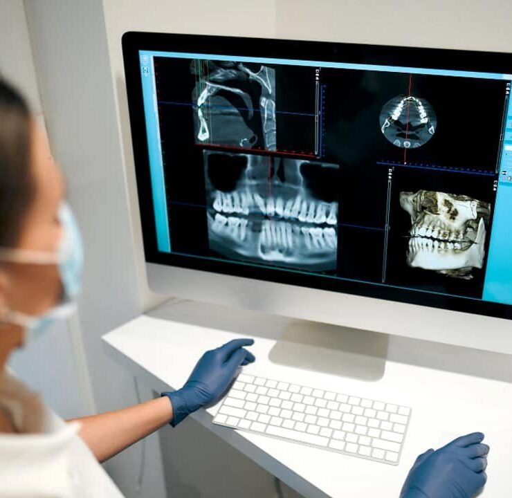
The Importance of Digital Imagery in Dentistry